Study Overview
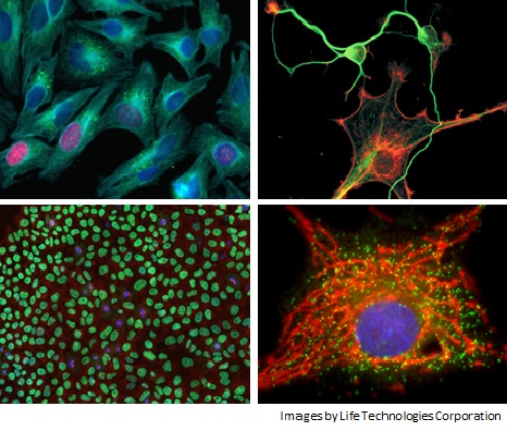
In this study, we use a fluorescence microscope to take pictures of cells and other samples and create easy-to-understand visual data that can also be used in promotional materials. We add test substances to cells and perform many kinds of evaluations, such as checking where target substances are located inside cells, observing changes in cells, measuring cell activity, studying how cells change into different cell types, and checking whether substances damage cells. We also offer time-lapse imaging to observe changes over time. Our experienced researchers can flexibly design test methods that match your study goals, so please feel free to contact us.
In addition, we have a wide range of equipment, including fluorescence plate readers and high-content screening systems. We also provide custom testing services that combine these different instruments.
Microscope used
List of Studies
| Study items | Staining Target・ Staining Method | Study purpose |
|---|---|---|
| Various immunostaining | Various targets | Verification of protein expression levels, cell differentiation observation, intracellular localization, etc. |
| DNA staining | DAPI | Verification of cell damage by nuclear staining of dead cells |
| Hoechst | Verification of cell damage by nuclear staining of live cells | |
| RNA staining | Newly synthesized RNA RNA-specific dyes etc. | Verify the effect of test substances on transcriptional activity |
| Organelle/ cell structure staining | Mitochondria | Verification of intracellular localization, and verification of mitochondrial activity and quantity |
| Endoplasmic reticulum | Verification of intracellular localization, etc. | |
| Golgi apparatus | Verification of intracellular localization and observation of morphogenetic changes | |
| Lysosomes | Verification of intracellular localization, etc. | |
| Cell membrane | Verification of intracellular localization and neurite outgrowth activity | |
| Adiposomes | Fat storage research using neutral fat staining, and verification of toxicity and side effects of test substances | |
| Intercellular junctions | Verification of cell-cell binding activity, including cell function activation and degradation | |
| Cytoskeleton | Actin, tubulin, etc. | Verification of intracellular localization and cytotoxicity of test substances |
| Living cells/ dead cells | Calcein AM | Verify the cytotoxicity and cell activation of test substances |
| Cell Tracker | ||
| BrdU staining | Verify cell division by staining cells during DNA synthesis | |
| Caspase Reagents | Verify the presence of apoptosis and necrosis | |
| TUNEL assay, etc. | ||
| Oxidative stress | Mitochondrial activity | Verification of oxidative stress by the presence or absence of reactive oxygen species generated from mitochondria |
| Nitric oxide | Verification of oxidative stress and signaling depending on the presence or absence of nitric oxide | |
| Reactive oxygen | Verification of antioxidant capacity based on the presence or absence of reactive oxygen species | |
| Thiol Reaction Reagents | Observation of intracellular redox state and verification of oxidative stress status | |
| Autophagy | Anti-LC3B staining, Lysosomal staining, etc. | Verification of the autophagosome formation promoting effect |
| Various ion concentration | Various ion indicators such as Ca2+ and H+ | Measurement of intracellular ion concentrations |
| Subcellular localization | GFP, YFP, RFP, etc. | Intracellular localization, intracellular movement/transport of various fluorescent fusion proteins |
*Studies will be mainly conducted by those with doctoral degrees.